By Ronnie LaCombe, PhD Candidate in Biological Sciences.
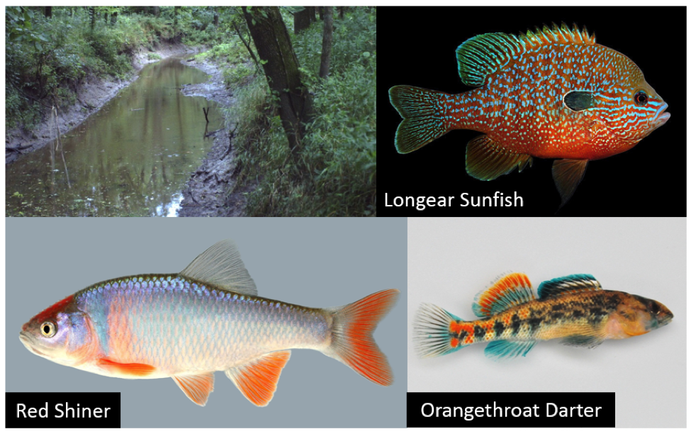
Ronnie has participated in events in Jefferson City, Union, and Fredericktown events
The human body is an amazing feat of nature. We are composed of trillions of individual cells, divided into countless specialized groups that form our organs, and all of them coordinate to keep us healthy. To do this, cells have developed many different methods of communicating. Sometimes they communicate over long distances, like how hormones work. You can think of this like sending a letter through the post office to be delivered to a friend out of state. Other times, neighboring cells talk to each other, kind of like passing a note to your friend in class. Biovisions at Harvard University produced one of my favorite videos about how cells work, and includes animates of cells communicating! You can find a shortened version here or a longer, narrated version here.
Cell communication can break down in several different ways. The message may never be sent, may be intercepted along the way, or may not be able to be received. When the systems break down, problems arise. Diabetes is an example of cell communication gone wrong. In type I diabetes, the insulin signal is never produced. In type II diabetes, the signal is reduced but cannot be received. Breakdown in cell-to-cell communication can also cause cancer, which is what my research focuses on.
I study a communication system that cells use to talk to their neighbors. I work with muscle cancer cells, called rhabdomyosarcoma (RMS) [rab-doh-mahy-oh-sahr-koh-muh]. It’s the most common soft tissue tumor in children, and unfortunately doesn’t have a specific treatment. Instead, it is treated with chemotherapy, surgery, and radiation, all off which have harmful side effects, so development of a targeted treatment is of high importance. You can find more information about RMS at focusonrhabdo.org, which includes background information, webinars, blog posts, and research papers among other information.
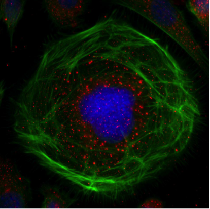
The communication system I study, the Eph receptors, is the largest family of its kind in humans, and is bound to the surface of cells. The signals they interact with are also surface bound, which means it’s a cell-to-cell contact communication system. We can visualize these proteins with a technique called immunofluorescence. In this technique, a tool specifically labels different parts of a cell, and is attached to a molecule that makes it glow when excited by light, just like glow-in-the-dark paint! This is what RMS cells look like with this technique:

In this cell, the blue is labeling DNA , the green is labeling cell structural components, and the red is labeling an Eph receptor.
I started my research by using immunofluorescence to detect how all of the Eph receptors are expressed in RMS in three species – dog, mouse, and human. If something is the same in all three species, it is likely important to RMS. While looking at this Eph screen, I stumbled upon an interesting observation – one of the Eph receptors was no longer located at the surface of the cells! Instead, it was on the inside of the cell in the nucleus, where your DNA is stored (where the blue is in the picture above). This means that it can no longer participate in cell-to-cell contact communication because it’s not on the outside of the cell anymore! So what is it doing there?
My project focuses on answering this question. There are a few things that the receptor could be doing on the inside of the cell. It could be interacting with DNA, it could be interacting with other proteins, or it could be doing both. By identifying what it is doing, we can learn about how it is contributing to RMS. It could be causing cancer cells to grow faster, causing them to spread (metastasize), or causing them to develop resistance to therapy, among other things. Once we know what it is doing, we hope to uncover an avenue for development of a new therapy.
A few people at the Jefferson City Science on Wheels event asked me about the videos I brought along with me – you can find a few videos here.
This is a publication from our lab that is open access, so anyone can see it! The videos are under the Supporting Information section.
Thanks for reading! If you have any other questions, you can e-mail me at vmlb88@mail.missouri.edu.